YOUR X-RAY. YOUR KNEE.
What’s My Tilt?
Your knee has its own unique angles and tilt, shaped by your thigh bone (femur), shin bone (tibia), and the contours of the joint surfaces where they meet. Together, these features help define your knee’s natural alignment.
JPG, PNG or DICOM
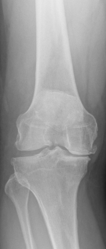
Start with your front-view X-ray.
Your X-ray
NO IMAGEPNG, JPEG or single-frame grayscale DICOM
Select a reference point to begin.
Which references are needed?
Hip: femoral head center. Knee: reviewed joint center used for this protocol’s hip–knee and knee–ankle axes. Ankle: ankle center. Femoral and tibial medial/lateral endpoints define each articular reference line. The common knee-center convention is recorded in the report and needs reproducibility validation.
Automatic preview EXPERIMENTAL
Research preview · proposals may be wrong. SGR is paused pending commercial permission. The original detector remains experimental and unvalidated.
Measurement tools · clinician use
Review the experimental proposals or mark the references manually. No review is arranged by this app.
Choose your evidence
Use a standing, full-length AP view with suitable rotation and positioning. For knee-only estimates, use a front-view image showing enough of both shafts to place two centers per bone. Mark the intended limb when both knees are shown. Unequal image stretching can distort angles.
How knee-only estimates work
Place two centers along each visible shaft, as far apart as the image supports, plus medial/lateral points on both joint lines. Confirm a suitable front view and correct point order. The femoral angle receives an assumed +5° correction; the tibial shaft is used as a mechanical-axis proxy. These assumptions are not patient-specific. Short or bowed shafts, rotation, flexion, implants and obscured landmarks can invalidate the estimate. Do not calculate when the required lines cannot be identified.
One 300-knee study reported 68.7% CPAK agreement with long-leg films. This app has not been clinically validated.
Short-film CPAK evidenceReview reference points
Joint-line points must represent the distal femoral and proximal tibial articular reference lines, not osteophyte tips. Automatic proposals can be incorrect; check every reference.
Enter mechanical angles from an existing reviewed measurement. These values are not measured from an image by this app.
Your alignment profile
NOT MEASUREDYour angles will appear here after measurement and review.
Your knee type CPAK
—Joint-line obliquity: —
Your CPAK type will appear after measurement.
What does CPAK mean?
Original CPAK: aHKA below −2° = varus; −2° through +2° = neutral; above +2° = valgus. JLO = MPTA + mLDFA: below 177° = apex distal; 177° through 183° = neutral; above 183° = apex proximal. Classification uses unrounded values.
aHKA estimates constitutional alignment and differs from the measured current mHKA. CPAK is a phenotype, not an arthritis grade, diagnosis, or treatment recommendation.
MacDessi et al., 2021 · original CPAK publication ↗